Interventional Echocardiography: A New Discipline in Cardiology
Prof. Dan Gilon, September 2016
Interventional cardiology continues to raise the bar, targeting new diseases, and developing interventions that require paradigm shifts. The first 3 decades of intervention concentrated on the treatment of coronary artery disease, with new approaches – specialized balloons, wires with coatings, bare-metal stents, drug-eluting stents and more.
Recently, we have begun to address structural heart disease in the cathlab. This group of heart problems deals with abnormalities of the valves and other structures of the heart. Until recently, treatment of narrowing of the aortic valve (the valve between the major pump – the left ventricle and the rest of the body) and especially leakage of the mitral valve (the valve between the left atrium which collects the blood from the lungs and the left ventricle) required open heart surgery. As patients grow older, they develop more medical challenges – many non-cardiac – which make cardiac surgical procedures more dangerous. In recent years, advances in imaging leading to improved understanding of anatomy, innovative approaches, and novel devices have made it possible to treat many of these valve abnormalities in the cathlab. These interventions began with a prosthetic stented valve that can repair the aortic valve that can be introduced into the heart in a cathlab procedure. More recently new devices have been developed to treat severe mitral regurgitation (insufficiency), also without the need for surgery. Devices to treat the tricuspid valve (between the right atrium and right ventricle) are under development.
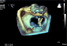
In order to deploy these devices properly, we have had to make advances in cardiac imaging as well, with real time 3 dimensional ultrasound taking on a crucial role. Patients require thorough examinations for diagnosis of their disease and the evaluation of the severity, and specific measurements to plan the procedure . During the procedure itself, echocardiography is needed to guide the procedure (as in placement of the device,) to evaluate the intermediate results, and in cases of procedural complications to diagnose and assess the so that life-saving interventions can be immediately performed. For example, during one type of procedure to repair the mitral valve, which uses the “Mitral Clip”, the echocardiographer assesses in advance whether or not the valve leaflets are suitable for clipping, and then guides the interventional catheterization procedure as to the introduction of the wires, the catheter that holds the clips and the precise placement of the “clips,’ catching the two leaflets of the insufficient valve and clipping them together, thus decreasing the leakage. The 3-dimensional capabilities of the echocardiogram done via a device in the patient’s esophagus is crucial. Without 3-D TEE such a procedure would be, if not impossible, much lengthier and much more dangerous for the patient.
Similarly, echo guidance is irreplaceable in several other structural interventions performed in the cathlab, including closure of holes in the heart (such as an atrial septal defect where there is a connection between the 2 atria (collecting chambers) of the heart), closure of the appendage of the left atrium in a patient with atrial fibrillation who cannot receive anticoagulation therapy as indicated (to prevent blood clots that may travel to the brain), and closure of leaks that develop around the edges of valves that have been surgically implanted.
There is now a new job description in the realm of cardiology- the interventional echocardiographer. This new specialty may not get all the glory, but it requires extensive knowledge, physical dexterity and nerves of steel. And without them, the great advances in the cath lab just wouldn’t happen.
Search
Recent Posts
Abuse and Heart Disease
Abuse and Heart Disease: Exposure to trauma and...The Fake News Fiasco
We are exposed to a tremendous amount of inform...Coaching For Change
The Pollin Center’s health coaching group is an...When on-line is off limits: Innovative Corona-tailored health promotion program for Ultra-orthodox women – Chevruta L’Chaim
Ultra-Orthodox (Haredi) women have worse health...21444
The stress, apprehension and the fears coupled ...

